
Did you know that 80% of animals over 5 years of age have some form of dental disease?
At VetsOne, we utilize the latest techniques and modern equipment
to provide the best dental care for your pet.
10 steps to a healthy mouth
- Physical examination. Every animal we see has an examination of the mouth performed, where possible, as part of the general physical examination. We can see if there is obvious disease in the mouth and we grade the severity of the dental disease from 1-4; with one being minor dental disease and 4 being major dental disease. This gives us an idea of what we may need to do during a dental procedure but it is difficult to fully examine the mouth of an awake pet. In addition, we can only see the crowns of the teeth, NOT the roots. Thorough inspection requires an examination under anaesthesia plus/minus radiography. Therefore, if dental disease is seen when we examine your pet, we will recommend anaesthesia and a COHAT (Comprehensive Oral Health Assessment and Treatment). We will provide an estimate for the procedures based on what we see in your pet during our consult exam but we may only become fully aware of the full extent of oral disease during the COHAT under general anaesthesia. It is also possible that mouth issues are not oral hygiene related, for example hidden tumors. Because they may be difficult to see in an awake animal, these are most often only discovered during the COHAT.
- Preoperative bloodwork and examination. Any animal that receives general anaesthesia at VetsOne gets a full pre-operative physical examination on the day of surgery. We also recommend blood tests are performed to make sure the animal is in good health. Whilst this is best practice for all animals, we view it as essential for all senior pets (those over 7 years of age).
- General anaesthesia. Dentistry requires an animal to be under a general anaesthetic so that we can properly examine the entire oral cavity and provide treatments pain free. The patient is anesthetized and IV catheter and anaesthetic monitors are placed. A qualified Veterinary Nurse closely monitors the patient during the dental procedure.
- Intraoral Radiology. We perform x-rays of the teeth for all patients undergoing a major dental procedure (Usually Grade 3 and 4) or for lower grades with any individual teeth that look suspicious. This is the only way to accurately evaluate the whole tooth.

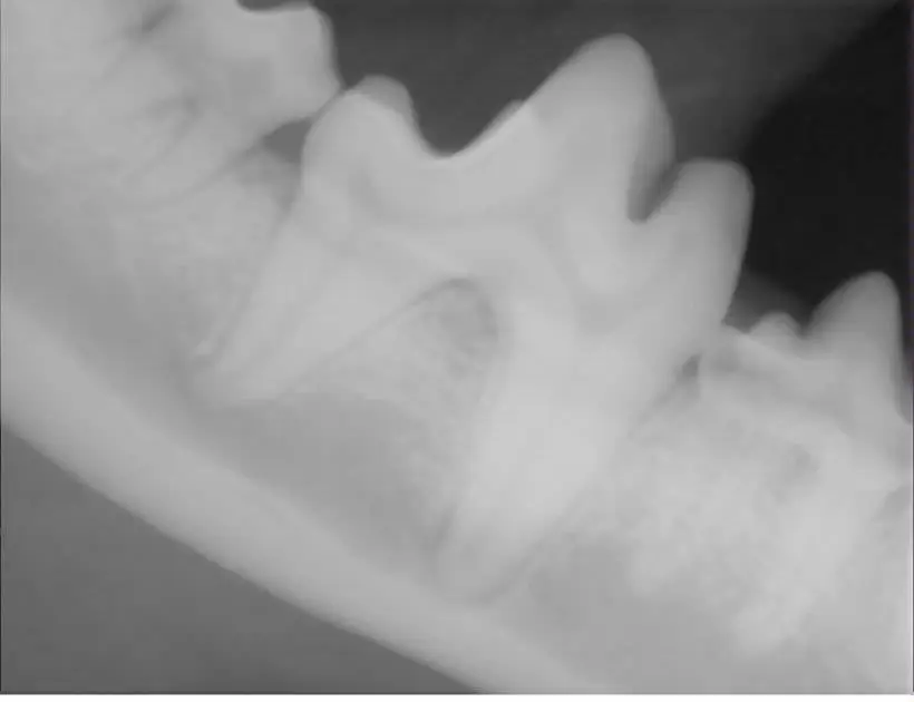
We use a digital dental x-ray system to evaluate each tooth.
The crown is the only portion of the tooth visible, because the root of the tooth is embedded in a socket in the jaw bone. In many cases the crown of the tooth may appear normal, but an x-ray of the tooth may reveal a problem with the root that requires treatment.
Here is an x-ray of a dog’s lower jaw (mandible) produced by our digital dental system.
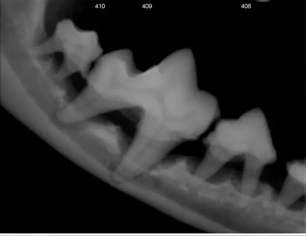
Once all of the teeth in the mouth have been x-rayed the treating veterinarian reviews the x-rays and decides on what therapy is required for each tooth.
This is an x-ray of a dog with severe dental disease. You can see that the bone around the roots of the teeth has receded away from the tooth roots. These teeth will need to be removed.

When we have removed the affected teeth we take another x-ray to make sure that all of the roots have been removed and there is no damage to the jaw bone.
5. Scaling. Scaling is the process where the tartar is removed from the teeth. Tartar is produced by bacteria that live on the teeth. Tartar causes inflammation of the gums (gingivitis) and this leads to recession of the gums, exposure of the tooth roots and eventually loss of the tooth. We remove the tartar with a combination of an ultrasonic scaler and hand scaling (just like the human dental hygienists). Removal of the tartar on the teeth is vital to improving the health of the mouth and it also removes the source of the patient’s halitosis (bad breath).
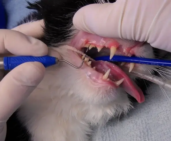
6. Periodontal probing. Once the teeth have been scaled the veterinarian examines each tooth individually with a periodontal probe. We use the probe to look for pockets. Pockets are caused by the gum losing its attachment to the tooth. Bacteria and tartar can accumulate in the pocket causing the wall of the tooth socket to erode. This leads to loosening of the tooth in the socket and eventually to tooth loss. A small pocket may be cleaned and flushed, but a deep pocket usually requires the affected tooth to be removed to avoid long-term problems.
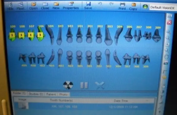
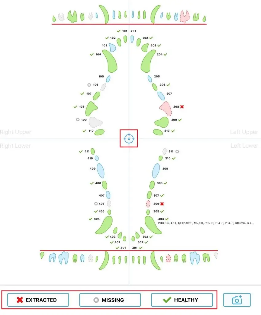
7. Charting. The combination of radiology and periodontal probing allows us to accurately diagnose any problems with the teeth and formulate a treatment plan. We use a digital charting system to record our findings and treatments.
This dental chart is used to accurately record findings and treatments.
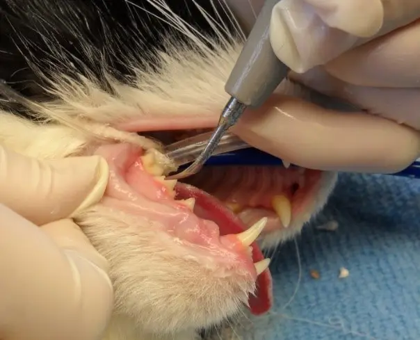
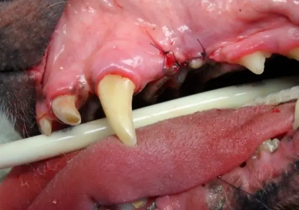
8. Extractions. If we decide that a tooth cannot be saved, it will be extracted. This becomes more likely in advanced dental disease (Grade 3 and 4) but individual teeth may have issues even at lower grades (for example tooth fractures. The first step is to place a local anaesthetic block to block the nerve. Even though the patient is under an anaesthetic removing a tooth can cause residual pain and local blocks offer the patient post-operative comfort. Once the block has taken effect, we elevate a flap of gum tissue to expose the bone. A high speed drill is used to cut the tooth into sections to allow for easier removal. The tooth is removed and then the socket is cleaned. A post extraction x-ray is taken to make sure that all of the roots have been removed. Finally, we close over the socket using the gum flap. This prevents food material from becoming lodged in the empty socket. The flap is sutured with a fine absorbable suture.
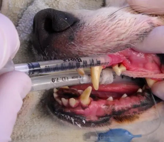
A local anaesthetic block is being placed prior to removal of the tooth with the exposed roots.
The tooth has been removed and a gingival flap is sutured over the socket.
9. Polishing. The final step to a routine COHAT is polishing the teeth. This helps remove any small lines in the teeth that may act as good attachment points for plaque and tartar.
10. Sealant. Once the scaling and treatment are completed we may apply a sealant to the teeth. This sealant helps to prevent bacteria from adhering to the teeth to slow down the accumulation of bacteria and tartar.
The importance of Post-operative care.
Firstly, we want your pet to recover quickly and comfortably, from what can be a major procedure (if extractions are involved). We will give specific post-operative instructions. This may include soft food, pain relief medication and no tooth brushing for a few days. We will also schedule a post-dental check with either a nurse or veterinarian, usually 3 days after your pet’s dental treatment. This check is complimentary.
Secondly, we want to reduce the amount of dental procedures your pet has during their lifetime. We know that plaque will start developing immediately after the teeth have been cleaned. Without a dental plan, it is likely your pet will need another procedure to clean the teeth within 6 months. It is vital that you have a home dental care plan to reduce tartar formation and reduce reliance on dental procedures. Please note though, that no treatment is perfect and can’t guarantee that procedures won’t be needed again – just that they won’t be needed as often.
We know there is a lot of information when you pick up your pet from us, so we will discuss home treatment options for you at the post-dental check.
Dental Consultations are FREE at any time, just phone us on 06 878 8666.
At VetsOne, your pet will receive the best personalized care tailored to their specific requirements. Our aim is that you and your pet go home with a smile.

Any questions or concerns please do not hesitate to have a chat with our team on 06 878 8666

